"Mars is not a dead planet -it undergoes climate changes that are even more pronounced than on Earth."
James Head of Brown University
The prevailing thinking is that Mars is a planet whose active climate has been confined to the distant past. About 3.5 billion years ago, the Red Planet had extensive flowing water and then fell quiet - deadly quiet. It didn't seem the climate had changed much since. Now, recent studies by scientists at Brown University show that Mars' climate has been much more dynamic than previously believed.
After examining stunning high-resolution images taken last year by the Mars Reconnaissance Orbiter, researchers have documented for the first time that ice packs at least 1 kilometer (0.6 miles) thick and perhaps 2.5 kilometers (1.6 miles) thick existed along Mars' mid-latitude belt as recently as 100 million years ago. In addition, the team believes other images tell them that glaciers flowed in localized areas in the last 10 to 100 million years - a blink of the eye in Mars's geological timeline.This evidence of recent activity means the Martian climate may change again and could bolster speculation about whether the Red Planet can, or did, support life."We've gone from seeing Mars as a dead planet for three-plus billion years to one that has been alive in recent times," said Jay Dickson, a research analyst in the Department of Geological Sciences at Brown and lead author. "[The finding] has changed our perspective from a planet that has been dry and dead to one that is icy and active."In fact, Dickson and his co-authors, James Head, a planetary geologist, and David Marchant, an associate professor at Boston University, believe the images show that Mars has gone through multiple Ice Ages - episodes in its recent past in which the planet's mid-latitudes were covered by glaciers that disappeared with changes in the Red Planet's obliquity, which changes the climate by altering the amount of sunlight falling on different areas.
NASA's Mars Global Surveyor and Mars Odyssey missions have provided evidence of a relatively recent ice age on Mars. In contrast to Earth's ice ages, a Martian ice age expands when the poles warm, and water vapor is transported toward lower latitudes. Martian ice ages wane when the poles cool and lock water into polar icecaps.The catalysts of ice ages on Mars appear to be much more extreme than the comparable drivers of climate change on Earth. Variations in the planet's orbit and tilt produce remarkable changes in the distribution of water ice from Polar Regions down to latitudes equivalent to Houston or Egypt. Researchers, using NASA spacecraft data and analogies to Earth's Antarctic Dry Valleys, reported their findings in the journal Nature."Of all the solar system planets, Mars has the climate most like that of Earth. Both are sensitive to small changes in orbital parameters," said planetary scientist Dr. James Head of Brown University. "Now we're seeing that Mars, like Earth, is in a period between ice ages," he said. This evidence of recent activity means the Martian climate may change again and could bolster speculation about whether the Red Planet can, or did, support life.Head and his team examined global patterns of landscape shapes and near-surface water ice Nasa's Mars orbiters mapped. They concluded a covering of water ice mixed with dust mantled the surface of Mars to latitudes as low as 30 degrees, and is degrading and retreating. By observing the small number of impact craters in those features and by backtracking the known patterns of changes in Mars' orbit and tilt, they estimated the most recent ice age occurred just 400 thousand to 2.1 million years ago.
Marchant, a glacial geologist who spent 17 field seasons in the Mars-like Antarctic Dry Valleys, said, "These extreme changes on Mars provide perspective for interpreting what we see on Earth. Landforms on Mars that appear to be related to climate changes help us calibrate and understand similar landforms on Earth. Furthermore, the range of microenvironments in the Antarctic Dry Valleys helps us read the Mars record."According to the researchers, during a Martian ice age, polar warming drives water vapor from polar ice into the atmosphere. The water comes back to ground at lower latitudes as deposits of frost or snow mixed generously with dust. This ice-rich mantle, a few meters thick, smooths the contours of the land. It locally develops a bumpy texture at human scales, resembling the surface of a basketball, and also seen in some Antarctic icy terrains. When ice at the top of the mantling layer sublimes back into the atmosphere, it leaves behind dust, which forms an insulating layer over remaining ice. On Earth, by contrast, ice ages are periods of polar cooling. The buildup of ice sheets draws water from liquid-water oceans, which Mars lacks.
Dickson and the other researchers focused on an area called Protonilus Mensae-Coloe Fossae. The region is located in Mars's mid-latitude and is marked by splotches of mesas, massifs and steep-walled valleys that separate the lowlands in the north from the highlands in the south.
The team looked in particular at a box canyon set in a low-lying plain. Images show the canyon has moraines - deposits of rocks that mark the limits of a glacier's advance or the path of its retreat. The rock deposit lines appear to show a glacier that flowed up the box canyon, which "physically cannot happen," Dickson said.Instead, the team deduced the ice in the surrounding plain grew higher than the canyon's walls and then flowed downward onto the top of the canyon, which had become the lowest point on the ice-laden terrain. The team calculated the ice pack must have been one kilometer thick by past measurements of height between the plain and the lip of the canyon. Based on the ice flow patterns, the ice pack could have reached 2.5 kilometers at peak thickness during a period known as the late Amazonian, the authors said.The finding could have implications for the life-on-Mars argument by strengthening the case for liquid water. Ice can melt two ways: by temperature or by pressure. As currently understood, the Martian climate is dominated by sublimation, the process by which solid substances are transformed directly to vapor. But ice packs can exert such strong pressure at the base to produce liquid water, which makes the thickness of past glaciers on its surface so intriguing.Dickson also looked at a lobe across the valley from the box canyon site. There, he saw a clear, semi-circular moraine that had spilled from an ancient tributary on to the surrounding plain. The lobe is superimposed on a past ice deposit and appears to be evidence of more recent glaciation. Although geologists can't date either event, the landscape appears to show at least two periods in which glaciation occurred, bolstering their theory that the Martian climate has undergone past Ice Ages.
Posted by Casey Kazan.
Related Galaxy posts:
Unraveling the Mysteries of Mars -Clues to Climate Change on Earth?Movie of NASA's Sites on Mars for Future Landings & Search for Ancient LifeMars Exploration: Secrets of the SoilIs There Life on Mars? NASA Goes Underground to Find OutNew Phoenix Mission Technology to Search for Mars LifeIs there an Interplanetary Mars-Earth Microbe Shuttle?"The Overview Effect": Is Space Travel Next Step in Human Evolution?Lonely Hearts of the Cosmos Revisited -NASA's Phoenix Probe & the Search for Extraterrestrial LifePhoenix Lander and the 'Canals' of Mars
Wednesday, May 21, 2008
Saturday, April 12, 2008
Nanoparticles hitchhike on red blood cells for drug delivery
By Society for Experimental Biology and Medicine, [RxPG] Researchers at the University of California, Santa Barbara have discovered that attaching polymeric nanoparticles to the surface of red blood cells dramatically increases the in vivo lifetime of the nanoparticles. The research, published in the July 07 issue of Experimental Biology and Medicine, could offer applications for the delivery of drugs and circulating bioreactors.Polymeric nanoparticles are excellent carriers for delivering drugs. They protect drugs from degradation until they reach their target and provide sustained release of drugs. Polymeric nanoparticles, however, suffer from one major limitation: they are quickly removed from the blood, sometimes in minutes, rendering them ineffective in delivering drugs.The research team, led by Samir Mitragotri, a professor of chemical engineering, and Elizabeth Chambers, a recent doctoral graduate, found that nanoparticles can be forced to remain in the circulation when attached to red blood cells. The particles eventually detach from the blood cells due to shear forces and cell-to-cell interactions, and are cleared from the system by the liver and spleen. Red blood cell circulation is not affected by attaching the nanoparticles."Attachment of polymeric nanoparticles to red blood cells combines the advantages of the long circulating lifetime of the red blood cell, and their abundance, with the robustness of polymeric nanoparticles," said Mitragotri. "Using red blood cells to extend the circulation time of the particles avoids the need to modify the surface chemistry of the entire particle, which offers the potential to attach chemicals to the exposed surface for targeting applications."The researchers have learned that particles adhered to red blood cells can escape phagocytosis because red blood cells have a knack for evading macrophages. Nanoparticles aren't the first to be piggybacking on red blood cells; the strategy has already been adopted by certain bacteria, such as hemobartonella, that adhere to RBCs and can remain in circulation for several weeks. The researchers say that it may be possible to keep the nanoparticles in circulation for a relatively long time, theoretically up to the circulation lifetime of a red blood cell - which is 120 days - if the binding between particles and the red blood cells is strengthened. The methodology is applicable to drugs that are effective while still attached to a red blood cell, although the researchers say that slow release from the red blood cell surface is also feasible.Mitragotri says "this mode of prolonging particle circulation has significant implications in drug delivery, potentially leading to new treatments for a broad variety of conditions such as cancer, blood clots and heart disease". Dr. Steven R. Goodman, Editor-in-Chief of the journal, said "this study dealing with the attachment of nanoparticles to red blood cells may also have important implications for future treatment of hematologic disorders. This fusion of modern nanobioscience with cell biology and hematology is precisely the type of interdisciplinary study that the new Experimental Biology and Medicine is interested in publishing."
digg_title = 'Nanoparticles hitchhike on red blood cells for drug delivery';
digg_bodytext = '"This mode of prolonging particle circulation has significant implications in drug delivery, potentially leading to new treatments for a broad variety of conditions such as cancer, blood clots and heart disease."';
digg_topic = 'health';
digg_skin = 'compact';
Thursday, February 14, 2008
LOOKING INSIDE THE HUMAN BODY USING POSITRONS
(August 2004)
PET or Positron Emission Tomography scans have become an important aspect of medical imaging and diagnoses, allowing doctors to look into the human body as never before. PET scans are different from other medical imaging techniques because they do not actually look at the body itself. Instead, PET scans look at bodily process by detecting the decay products from radioactive tracers injected into the body. Radioactive tracers are designed to mimic naturally occurring substances and tend to deliver less radiation than an X-Ray.
When the radioactive tracers decay inside the body, they release a positron, the antimatter equivalent of electrons. When the positron encounters one of the billions and billions of electrons inside your body, it annihilates in a flash of light called gamma rays-it is these gamma rays that are the positron emissions which PET scanners detect. The donut ring of the PET scanner is lined with gamma ray detectors.
Alternative Medical Imaging Techniques
There are many types of medical imaging techniques available to doctors and patients. Each techniques use slightly different technologies, allowing the doctor to focus on different aspects of the human body.
The simplest and most common technique used to look into the human body is the X-Ray. X-Rays are high-powered beams of light that can pass through some parts of the human body, like skin, but are stopped by other parts, like bone. Using X-Rays to expose film (much the same way that visible light exposes film in a camera) allows doctors to examine the skeletal structure with ease.
CT or CAT, Computerized Axial Tomography, scans also use X-rays but in a slightly more complicated way. A CAT scanner consists of a donut shaped ring containing numerous X-Ray tubes that shoot out beams of X-Rays. Patients slide through the donut hole while X-Ray detectors that are also around the donut ring record the X-Rays after they have traveled through the body. The computerized component of CAT scans involves interpreting all the X-Ray signals and combing them into a coherent image. Because CAT scanners only take X-Ray pictures of your body in thin slices, computers are needed to combine each thin slice into a comprehensive three-dimensional image. This technique of looking at the body in thin slices is called Tomography. CAT scans image the body in slices perpendicular to the axis stretching from the feet to the head. These slices fall in the axial plane of the body.
Ultrasound imaging uses a completely different technology than X-Rays. An Ultrasound probe emits ultrasonic sound waves, sounds above the range of human hearing, into the body. While most of these ultrasonic sound waves travel straight through the body, a small amount reflect off the transition layers between different types of tissue. The echoes from these transmission layers are picked-up by the Ultrasound probe, fed into a computer and then rendered into a two-dimensional image. Ultrasound technology uses sound echoes to look into the human body much the same way bats and submarines use sound echoes to see.
MRI, or Magnetic Resonance Imaging, uses magnetic fields to peer into the human body. The MRI machine contains a very large magnet that surrounds the patient with an intense magnetic field. When patients are immersed inside this magnetic field all of the billions and billions of hydrogen atoms inside their body align with it. Inside the MRI machine, radio frequency pulses are applied to specific parts of the body to excite some of these hydrogen atoms. Images are created based on how the exited hydrogen atoms lose their energy. These images are fed into a computer to generate an incredibly detailed look inside the human body.
How is PET different?
PET scans are different from all the other types of medical scans because they never actually look at the human body itself. Instead, PET scans use radioactive chemicals to look at different processes inside the human body. The actual PET scan machines are similar to CAT scan machines as they are both donut shaped and use axial tomography. But unlike CAT scanners, the donut ring of PET scanners is lined only with detectors. These detectors are used to pick up positron emissions.
But what on earth is a positron? The simplest answer is that a positron is an anti-electron. So what is an anti-electron? There are billions and billions of electrons in your body. They are the negatively charged parts of atoms which orbit the positively charged nucleus. Every one of your atoms has electrons and electrons are considered to be what physicists call matter. Positrons, or anti-electrons, are antimatter-having many of the same properties as matter but with opposite electric charges. An electron is negatively charged while the positron is positively charged (electrons are sometimes called negatrons to differentiate them from positrons).
The important thing about positrons is that when they encounter electrons, they annihilate in bursts of light called gamma rays. These gamma rays are the positron emissions that the PET scanners detect.
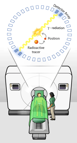
Before undergoing a PET scan, patients are given radioactive tracers. When these radioactive tracers decay, they emit positrons that soon annihilate with one of the billions of electrons inside your body to produce gamma rays. These gamma rays are always emitted back-to-back, or in the opposite direction from each other. This allows PET scanners to extrapolate where in the body the gamma rays originated.
Radioactive Tracers and PET Scans
What makes PET scans so unique is that they can be used to look at a number of physiological processes taking place inside the human body. Specific radioactive tracers allow doctors to study different biological processes. The radioactive tracers that PET scans use are manufactured to mimic naturally occurring substances already used by the body. As the body incorporates the radioactive tracers into its systems, the PET scan can monitor their progress and examine the specific bodily processes that use the tracer. Radioactive tracers are designed to deliver a minimum amount of radiation to the patient, often less than would normally be received in an X-Ray.
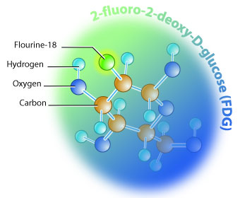
Worldwide, 95 % of PET scans use a tracer known as FDG, which is a fluorine-18 atom attached to a glucose molecule. FDG scans are used to detect and examine cancerous tumors throughout the body. FDG scans can also be used to study the fine detail of sugar metabolism in the brain. At TRIUMF and the University of British Columbia Hospital, the PET scan group focuses specifically on brain activity.
There are a variety of radioactive tracers that allow PET scanners to examine different aspects of brain function. One example is the Oxygen-15 radioisotope, used to study oxygen metabolism in the brain. Oxygen-15 can also be attached to carbon monoxide for the study of blood volume, or to water for the study of blood flow in the brain. There are hundreds of radioactive tracers available for PET scans, however, only several dozen are commonly used.
The Cyclotron and Rabbit Hole
To create these radioactive tracers, doctors and technicians make use of a particle accelerator called a cyclotron. Cyclotrons are high-tech machines used typically in nuclear and particle physics.
Radioactive tracers are designed to decay relatively rapidly so that there is little delay between the time that they are ingested and when they start to produce a measurable signal. Carbon-11, for example, has a half-life of only 20.3 minutes. Most PET scan groups do not have a cyclotron located close enough to create and deliver enough radioactive tracers before to many of them decay. Fortunately for the University of British Columbia Hospital and the PET scan group, their cyclotron is located at TRIUMF, only 2.4 kilometers away. Yet even though TRIUMF and the UBC Hospital are so close, they are still too far apart to package and drive the radioactive tracers between them.
To solve this dilemma, TRIUMF at UBC hospital scientists have created the world’s longest rabbit hole. This rabbit hole is a 2.4 kilometer long pneumatic pipeline that connects the cyclotron at TRIUMF to the PET scanner at the UBC Hospital. The radioactive tracers are dropped into the pipeline at one end and hop out the other. The rabbit hole allows PET scientists to make as much use of their cyclotron as possible.
UBC PET Scan Research
The PET scan program at TRIUMF and UBC is currently focusing on understanding the brain activity of Parkinson patients. Researchers are focusing on how the origin, progression, therapy and complications of therapy of Parkinson’s disease affect the brain. Researchers hope to better understand the effects and progression of the disease in order to promote a cure.
As well, the UBC PET group is constantly looking for new radioactive tracers to help improve the PET scanning technique and process. Combined with the expertise at TRIUMF, the UBC PET scan group offers unique opportunities in PET scan imaging.
Friday, January 11, 2008
Prof. L.K. Dadhich -hats off to you

Professor L.K. Dadhich
Environmentalist
Professor Laxmi Kant Dadhich is the green man of India and combines in his person an Environmentalist, Botanist, Ecologist, and an Information Technologist. He is an M.Sc. in Botany, M.A.D.E. in Distance Education, B.J.M.C. in Information Technology, Ph.D. in Ecology and Environmental Science and is an orator par-excellent. He has thirty-two years' research and teaching experience. He is an academic counselor too. He has been writings scripts for Radio and TV. He has chaired the technical sessions related to multimedia technology in distance education organised by UNESCO and IGNOU.He has conducted EIS for setting up industries. He was commissioned officer as Squadron Leader in the Indian Air Force (till 1999). He trained the youth in environmental conservation, education and global scenario and taught on topics related to Aviation. He has since retired from the position. He was the Senior Coordinator for a project sponsored by the Ministry of Environment and Forests, Govt. of India on Environmental Awareness. He was the Senior Advisor to a film on Environment produced by the Reyerson, Canada. He was a Consultant with a film 'Greening the Desert' produced by the Indian Army and TV Centre, Govt. of India. He also acted as the Consultant with Multimedia Technology & Distance Education. In Singapore, he worked on Environment management plans at a Seminar/Workshop organised by IUCN and IES; in Nepal he acted as a Research Coordinator of a Workshop 'Environment Education and Attitudes towards Environment Conservation in Developing Countries' organised by ISTE. For the last 33 years he has been working closely with several research centres and universities on issues of EIA, EIS, environment protection, water quality, pollution and other related matters, and in particular he has been having exchange relations with members from ISTE, Varanasi; JAC, New Delhi; GEAG, Gorakhpur;CEE, Ahmedabad; and CSE, New Delhi. He has been providing communication consultancy in a regular manner to UNEP, ELCI, Nairobi, Kenya. He has been a Fellow of the United Writers Association of India(FUWAI), Chennai, and Life Member Fellow of the National Institute of Ecology, New Delhi.
His biographical sketches were published in Reference Asia, Indo-Arab Who's Who, Indo-America Who's Who, Indo-European Who's Who, Learned Asia Who's Who, Asia-Pacific Who's Who, and 20th Century Admirable Achievers. He has published 37 research papers in journals and books of international repute and authored 11 books/reports, besides giving talks/programs telecast/broadcast by the AIR/TV. He has supervised 13 dissertations/project works/Ph.D. theses.
He was bestowed upon the WEC-IIEE-IAEWP International Award by the President of India for his concept of environmental citizenship; award for Environment Protection and Conservation by the C.M., Rajasthan; Swarn Puraskar by ISCD, Rohtak, Haryana; award for imparting training to the youth for Environmental Conservation by the Environment Minister, Rajasthan; Bharat Mata award by Astro Research project, Kolkata; award for forest extension activities in Kota Distt. by the Forest Minister, Rajasthan. He received Commendations from the Director-General of NCC and from the District authorities for his work in the field of environment and youth leadership training. Honours were conferred upon him by various organisations like TEEJ, Lions Club, Thalassemia Society, Blood Bank Society, etc.
He is the member of Environment Liaison Centre International, Nairobi, Kenya; Global Forum for NGO's for Natural Disaster Reduction (GFNDR) Yokohama, Tokyo, Japan; International Society for Tropical Ecology, Varanasi; Gorakhpur Environment Action Group, Gorakhpur; National Institute of Ecology, New Delhi; Centre for Science and Environment , New Delhi; Technocrat Council of India, Kota; Environment Society (Paryavaran Parishad), Kota; Society for Eco-Balance, Bhopal ; Joint Assistance Centre, New Delhi;and Paryavaran Vahini, Ministry of Environment and Forests, New Delhi.
The citizens of India and the world at large is proud of having such a multi-dimensional personality among them. Naturally, he has become a role-model for the young men and women aspiring to lead a meaningful life while remaining closer to the mother nature. All of us wish him a long and healthy life and should learn the meaning of an ideal life while sitting at his feet.
S.C. Scientists Find New Shark Species
The Associated Press
Monday, June 12, 2006; 11:26 PM
COLUMBIA, S.C. -- A new genetically distinct species of hammerhead shark, the ninth recognized species of hammerhead, has been discovered off the South Carolina coast, scientists say.
The new species appears to be rare and lives off the South Carolina coast. Classified under the genus sphyrna, will be called the "cryptic species" for the time being.
 This is an undated photo provided by the University of South Carolina Media Relations, shows a new species of hammerhead shark found off the coast of South Carolina. Joe Quattro, a biology professor at the Univerity of South Carolina, worked with Jim Grady of the University of New Orleans and Trey Driggers of the Natioal Marine Fisheries Service in making the find. (AP Photo/Univeristy of South Carolina) (AP)
Save & Share | |||||||||||||||||||||||||

Joe Quattro, a biology professor at the University of South Carolina, worked with Jim Grady of the University of New Orleans and Trey Driggers of the National Marine Fisheries Service in making the find.
Quattro discovered the new species while studying along the coast with biologists from the South Carolina Department of Natural Resources.
Quattro and his colleagues found that genes in the mitochondrial DNA _ the DNA passed from mother sharks to their offspring _ differed significantly among sharks that were classified as scalloped hammerhead sharks.
The studies also revealed that another independent genetic marker differed substantially between the two groups of scalloped hammerheads.
"This cryptic shark was genetically distinct," said Quattro, whose research was published recently in the journal, Marine Biology.
Scalloped hammerheads are common along the coast and sharks of the cryptic species were found from Florida to North Carolina. The newborn cryptic sharks, however, were found mainly along the South Carolina coast.
"The apparent abundance of the cryptic species in coastal South Carolina could be a result of sampling, but it might also highlight the fact that the South Carolina bays are the more important nursery grounds for the cryptic species," Quattro said.
Something as simple as the salinity of the water might explain why the sharks prefer the South Carolina coast, said Quattro, who plans a field trip this summer to tag the cryptic sharks so scientists can learn more about them.
Because they seem to have a narrow geographic distribution, the sharks may be at greater risk for extinction.
"If South Carolina's waters are the primary nursery grounds for the cryptic species and females gather here to reproduce, these areas should be conservation priorities," Quattro said.
Tuesday, January 8, 2008
Foods that Heal
We've all heard about the protective power of food. Some foods like fruits and vegetables are good for us and some elements of our diet, like too much fat, are bad for us. At Enhancing Foods to Protect Health, a new research center in Indiana, scientists are working to perfect food that may one day be designed based on an individual's health needs.
Excess fat consumption has long been the bane of those seeking to be healthy but fats do play beneficial roles in the body. Generally speaking, based on the common diet in the United States, too much omega-6 fatty acids are consumed and too little omega-3 fatty acids. Omega-6 fatty acids are common in corn and soy oils while omega-3 fatty acids are common in canola and fish oils. This is crucial, as the ratio of omega-6 fatty acids to omega-3 fatty acids should be in the range of 4 to 1 up to 10 to 1.
Diets in the U.S. have shifted such that the ratio is around 25 to 1. With such a high ratio, the risk for some types of cancer and arteriosclerosis increase dramatically.
The researchers at the Center are exploring ways to produce so called "designer eggs" with a better-balanced ratio of omega-6 fatty acids to omega-3 fatty acids.
Purdue food science Professor Bruce Watkins and graduate student Amy Devitt check the color and texture of eggs that have a better-balanced fat content.
In the study, hens were fed supplemental conjugated linoleic acid (CLA) in an attempt to see if the eggs produced by the hens would contain a better balance of fatty acids. And indeed, those hens fed the supplemental CLA produced eggs with a higher level of CLA than hens that were not fed the CLA.
The researchers then fed the hens a blend of two fatty acids. The hens were fed a combination of docosahexaenoic acid (an omega-3 fatty acid) and the conjugated linoleic acid. Again, the levels of the beneficial fatty acids were higher in those hens fed the supplements versus those who were not given the supplements.
The researchers hope that these studies will lead to the development of foods that have a higher ratio of beneficial fatty acids to nonbeneficial fatty acids.
Work still remains to be done as the eggs produced by the hens had a somewhat tough yolk and a very different texture from traditional eggs. The researchers are optimistic that with fine-tuning of the diet of the hens, an egg with the proper fat balance as well as a good taste could be produced.
What do you think? How might "designer foods" decrease the risk of heart disease and cancer? Do you think this is the best way to reduce risk or are there better ways? Come on over to the Biology Forum to share your thoughts, feelings, and opinions. 'Til next time...
Brain Power
Researchers at Duke University and MCP Hahnemann University have developed a technique for using brain signals to control a robotic arm. This feat was accomplished by recording signals from electrodes that were implanted in the brains of rats. It is believed that this new method may some day offer hope to those suffering from spinal cord injuries who have prosthetic limbs. Theoretically, the electrodes could be implanted into the brain to allow the person to have control over limb movement, much as they would an actual limb. In the experiment, rats were taught to operate a robotic arm by pressing a lever. Pressing the lever resulted in the rats receiving a reward. Researchers recorded the neuronal activity responsible for muscle movement using implanted electrode arrays. Once the specific groups of neurons that were used for muscle movement when pressing the lever were identified, researchers changed the control of the robotic arm from the lever to the electrode implants. The rats promptly learned that they could move the robotic arm to receive a reward without having to press the lever. All they had to do was to activate the particular neurons in the brain that they had previously used when physically pressing the lever. This ground-breaking study established the first tangible evidence that neuron signals can be used to control external devises. Prior to this study, scientists suspected that neuronal control of external devices was possible but there was no demonstrable proof. Researchers speculate that the knowledge gained from this study could be used to develop new techniques to treat those suffering from a variety of disabilities including spinal cord injuries, cerebral palsy, and locked-in syndrome. Since those with locked-in syndrome may have intact thinking skills but no ability to interact with their environment, external control of devices through neurons may be particularly helpful. The researchers also strongly emphasize that there are still significant technical obstacles that must be overcome before any attempts at human clinical trials can begin. They do however believe that these obstacles are not impossible to overcome.
Thursday, January 3, 2008
Wildfacts
Wildfacts
|
| ||||||||||||||||||||||||||||||||||||||||||||||||||||||||||||||||||||||||||||||||||||||||||||||||||||||||||||||||||||||||||


Subscribe to:
Comments (Atom)